Monday, July 21, 2008

AFP

In adults, high blood levels (over 500 nanograms/milliliter) of AFP are seen in only three situations:

  • HCC
  • Germ cell tumors (cancer of the testes and ovaries)
  • Metastatic cancer in the liver (originating in other organs)

Several assays (tests) for measuring AFP are available. Generally, normal levels of AFP are below 10 ng/ml. Moderate levels of AFP (even almost up to 500 ng/ml) can be seen in patients with chronic hepatitis. Moreover, many patients with various types of acute and chronic liver diseases without documentable HCC can have mild or even moderate elevations of AFP.

The sensitivity of AFP for HCC is about 60%. In other words, an elevated AFP blood test is seen in about 60% of HCC patients. That leaves 40% of patients with HCC who have normal AFP levels. Therefore, a normal AFP does not exclude HCC. Also, as noted above, an abnormal AFP does not mean that a patient has HCC. It is important to note, however, that patients with cirrhosis and an abnormal AFP, despite having no documentable HCC, still are at very high risk of developing HCC. Thus, any patient with cirrhosis and an elevated AFP, particularly with steadily rising blood levels, will either most likely develop HCC or actually already have an undiscovered HCC.

An AFP greater than 500 ng/ml is very suggestive of HCC. In fact, the blood level of AFP loosely relates to (correlates with) the size of the HCC. Finally, in patients with HCC and abnormal AFP levels, the AFP may be used as a marker of response to treatment. For example, an elevated AFP is expected to fall to normal in a patient whose HCC is successfully removed surgically (resected).

There are a number of other HCC tumor markers that currently are research tools and not generally available. These include des-gamma-carboxyprothrombin (DCP), a variant of the gamma-glutamyltransferase enzymes, and variants of other enzymes (e.g., alpha-L-fucosidase), which are produced by normal liver cells. (Enzymes are proteins that speed up biochemical reactions.) Potentially, these blood tests, used in conjunction with AFP, could be very helpful in diagnosing more cases of HCC than with AFP alone.

portal vein thrombosis./ obstruction

Pathophysiology

The portal vein forms at the junction of the splenic vein and the superior mesenteric vein behind the pancreatic head, and it can become thrombosed or obstructed at any point along its course. In cirrhosis and hepatic malignancies, the thromboses usually begin intrahepatically and spread to the extrahepatic portal vein. In most other etiologies, the thromboses usually start at the site of origin of the portal vein. Occasionally, thrombosis of the splenic vein propagates to the portal vein, most often resulting from an adjacent inflammatory process such as chronic pancreatitis.

Inherited and acquired disorders of the coagulation pathway are frequent causes of portal vein thrombosis. Inherited disorders include mutations in the prothrombin gene G20210A as well as deficiencies of various intrinsic anticoagulation factors, such as protein C and protein S, and activated protein C resistance. Acquired disorders include antithrombin III deficiency resulting from malnutrition, sepsis, disseminated intravascular coagulation, inflammatory bowel disease, liver disease, or estrogen use.

Stasis can be another major category for portal vein thrombosis. The global resistance to hepatic blood flow produced by cirrhosis is a common cause. Sclerotherapy for esophageal varices has been postulated as a possible mechanism though not proven thus far. The portal vein or its tributaries can be obstructed by adjacent tumor compression or invasion. Infectious and inflammatory processes may also lead to venous thrombosis.

Portal vein obstruction does not affect liver function unless the patient has an underlying liver disease such as cirrhosis. This is partially due to a rapid arterial buffer response, with compensatory increased flow of the hepatic artery maintaining the total hepatic blood flow. Formation of collaterals occurs rather rapidly as well, and they have been described as early as 12 days after acute thrombosis, though the average time to formation is approximately 5 weeks.

The development of a collateral circulation, with its attendant risk of variceal hemorrhage, is responsible for most of the complications and is the most common manifestation of portal vein obstruction. Other sequelae of the subsequent portal hypertension, such as ascites, are less frequent. Rarely, the thrombosis extends from the portal vein to the mesenteric arcades, leading to bowel ischemia and infarction.

portal vein obstruction

    • causes-
    • In adults, cirrhosis is the major etiology, accounting for 24-32% of cases of portal vein thrombosis.
    • Neoplasms are another major cause, accounting for 21-24% of cases of portal vein obstruction, with hepatocellular carcinoma and pancreatic carcinoma causing most of these cases. These tumors can cause compression or direct invasion of the portal vein and lead to thrombosis by inducing a hypercoagulable state. Local ablative therapies for hepatocellular or metastatic disease have been linked to its development.
    • Although less common than in children, infections (predominantly intra-abdominal) still play an important role, with a particular association to Bacteroides fragilis bacteremia.
    • Myeloproliferative disorders and inherited or acquired coagulation disorders account for 10-12% of cases in adults.
    • Approximately 8-15% of cases have been reported to be idiopathic in the recent literature. For other less common etiologies, such as abdominal trauma, surgery, and inflammatory bowel disease

      Physical findings-

      • Splenomegaly is found in 75-100% of patients, most presenting in the chronic stage. Mild hepatomegaly is often present, as is right upper quadrant epigastric tenderness, especially in the acute setting.
      • Ascites is found infrequently. Stigmata of chronic liver disease, such as spider angiomata or palmar erythema, are usually found in the presence of underlying liver disease.
      • The presence of caput medusae indicates posthepatic or intrahepatic portal hypertension because it forms by recanalization of the umbilical vein, which connects with the left hepatic branch of the portal vein. It should not be observed in isolated extrahepatic portal vein obstruction because the obstruction is below the origin of the umbilical vein.
      • In children, growth retardation may be present.
      • Abnormalities of the extrahepatic biliary tree may occur in 80% of cases due to compression by choledochal or periportal varices or from ischemic stricturing. These findings manifest by jaundice, cholangitis, hemobilia, cholecystitis, or a hilar mass that can be mistaken for a cholangiocarcinoma.


      ,

renal failure

Q: What do you think is the cause of renal failure
A:Being of Asian descent Diabetes is highest on my list but could also be due to hypertension, glomerulonephritis, adult polycystic kidney dse, nephrocalcinosis

Skin

Skin

A straw-yellow hue is found in cancer cachexia.

Paleness may be from anemia, dysemia, leukemia, amyloid degeneration, or Bright's disease.

Articular rheumatism is marked by paleness, and profuse sweats with strong acid odor.

Anger, fear, and jealousy cause paleness. The cause is vascular spasm. Fainting causes pallor.

Plethoric people are too red in color. A florid complexion means the sanguinous temperament and does not mean too much blood.

Unconsciousness may be from syncope (fainting). The face is pale; either no pulse or very light; the breathing very low and quiet. There are no signs of distress; the face is usually composed.

2. Appearance Of Patient

2. Appearance Of Patient

The patient's appearance will tell whether or not he is able to meet the requirements of existence. He looks able to carry on his work--his particular occupation- or he does not. If he does not, he will give the appearance of being sick with either acute or chronic disease.

At the bedside the patient may look robust, sick, collapsed, bluish or cyanosed, thin, fat, with thick and short neck, or long and slender; he is on his back with legs extended, or with the legs drawn up; or on the side with legs drawn up against the abdomen.

The patient may be unable to give a history or describe his symptoms.
Decubitus (Lying Down)

The manner of lying is significant. On the back means exhaustion. This is the position when a patient has lost consciousness.

In a faint or anemia of the brain, the head drops; in congestion of the brain, the head must be supported on several pillows; in asthma of the lungs, bronchi, or caused by the heart, the patient must have much pillow support.

In heart disease the patient lies upon the right side. A normal person can lie on either side equally well.

When heart disease is advancing to the fatal state, the position is sitting, with head and shoulders supported by pillows.

Pain in the abdomen will cause the sufferer to press upon it, or lie on a pillow. Pressure gives some relief. When the pain is intense there will be twisting and writhing.

In peritonitis, appendicitis, cystitis, gallstones, cancer of the stomach and bowels, the tendency is to draw the legs on the abdomen. In peritonitis, the patient will usually be on the back, with legs drawn up.

In gastric ulcer, when suffering with pain, if the ulcer is in the front wall of the stomach, the patient will lie on his back; if the posterior wall is the location of the ulcer, the patient's position will be lying on the abdomen; or upon the right or left side, if the disease is of the right or left side. These positions relieve pressure on the ulcer.

In tubercular meningitis, the child lies on the side, with legs strongly drawn up against the thighs.

Facial Expressions

Facial Expressions

Disease as expressed in the face and posture.

Facies cardiac (heart): An anxious expression seen in the early stages of chronic valvular disease.

A purple or bluish appearance of the face, especially about the eyes, temples, and ears, with veins showing on the nose and sometimes on the cheeks, intensified by lying down: Caused by high blood pressure and an approaching dangerously plethoric state of the body.

Hepatic face: An earthy appearance; yellow tinge, jaundice.

Hippocratic face: Indicating rapid approach of death--pinched nose; hollow temples; eyes sunken; ears leaden and cold; lips relaxed; skin livid, and if the skin is pinched it returns slowly to the plane from which it was pinched or drawn.

Ovarian face: Features emaciated and sunken; anxious expression; forehead furrowed; eyes hollowed; nostrils open and sharply drawn; lips full and compressed; angles of mouth drawn and wrinkled, puckered but protruding"fish mouth."

The stupid face is that of typhoid.

Gastric face in children: A white line around the mouth, extending up by the side of the nose, shows irritation from improper feeding. Add to this sign pungent breath and vomiting, and the child has gastritis.

Gastric face in adults: Chronic irritation of the stomach in adults is indicated by a dragging-down of the comers of the mouth. Add to this drooling or driveling of saliva, and the indication is of starch poisoning; and if there is a broad, pallid tongue, the evidence is strong for overeating on starch.

Hysteria is marked by staring and an ecstatic expression.

Epilepsy is marked by a stupid face after an attack.

Protruding eyes and expressionless face in Graves' disease.

They lypermaniac has sadness written in his face. In general paralysis the countenance is composed and satisfied. The enebriate has trembling bps and a wandering expression.

The child with enlarged tonsils and adenoid growths has a stupid expression; the mouth is open, the lips hanging; the nose is expressionless.

The red nose, enlarged veins, bluish lips, cyanosed cheeks, and puffiness of face of the drinking man are called the mitral face. Where the aorta is diseased there is intense pallor. In Bright's disease the face is swollen and white.

The signs of croup are well known, but the type of disease is not so easily told. There are coughing and suffocating when a foreign body is in the air-passage.

Expiratory disturbance is marked by flushed face, puffed and bluish; the eyes are suffused, and the veins stand out.

In marasmus the features are drawn, the furrows deepened, the neck hollow; emaciation is marked, and, when profound, the whole appearance is that of the monkey.

The consumptive appearance is that of emaciation; protruding, flushed cheeks; pinched nose, with flaring nostrils; short, quick, jerky breathing; halting speech, and more or less suppressed voice.

When the face looks smaller--shrunken--and the nose is thin, long, and drawn, the bones prominent, the skin pale and covered with cold sweat, and, when drawn or pinched, the fold remains for some time, this is the facies of peritonitis, intestinal obstruction, renal and hepatic colic.

Fainting: The heart stops; the patient turns pale and falls motionless, but there is no distortion of the face; breathing is suspended.

Apoplexy: The patient is motionless and lies on the back; all animation is suspended; only breathing and pulse continue; the breathing is noisy, and gradually grows more stertorous. If the patient does not react and improve, the breathing and heart action gradually decline, the skin becomes drawn, the nose thinner and longer, the eyes dull, partially closed, glassy. The breathing stops, starts and continues, until it finally ends with a slight bodily convulsive movement.

Physical appearance must be noted--all deviations from the normal mean something.

Deformities, such as rickets, shorten the stature and cause the head to appear too large; the spine is incurved, the pelvis is deformed, the limbs are curved, the ribs project forward.

When the muscles become atrophied they cause general deformity.

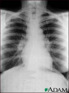
Alterations of the heart or lungs cause deformities of the chest.

The bowels are often too large and distended from gas, fat, or ascites; in fevers, from tympanitis and inflammations.

Enlargement of the liver or spleen causes a large abdomen in the upper region; in the lower abdomen, enlargement may come from tumors, distended bladder, or a gravid uterus.

A large swelling at the base of the great toe, with the toe pointing outward, indicates a bunion. This deformity usually means that there is a slight rheumatism. Deformity of the third joint of the fingers--nodes of Heberden--means arthritis deformans. The nodes of Bouchard on the second joints of the fingers indicate dilation of the stomach--a disturbed nutrition from overeating of the carbohydrate foods. Joint distortions indicate gout, rheumatism, or injury; not infrequently they mean all of these. Frequently injury is complicated by rheumatism.

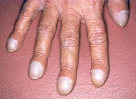
Hippocratic fingers (clubbing of finger-tips, with incurving nails) indicate heart or lung disease--scrofulous diathesis.

face,mrcp

Sunday, July 20, 2008

Synovial Fluid Analysis

Synovial Fluid Analysis

Print This Page
E-mail This Page

The most important reason for performing joint fluid analysis is to rule out septic arthritis. Synovial fluid analysis can demonstrate local inflammatory response, infection and the presence of crystals. Synovial fluid effusions are classified into five general etiologic categories: noninflammatory responses, inflammation, sepsis, crystal induced and hemorrhage. The most common diseases associated with each category are summarized in the following table.


Effusion

Diseases

Noninflammatory

osteoarthritis, trauma, osteochondritis, pigmented villonodular synovitis, sickle cell disease, neuropathic

Inflammation

rheumatoid arthritis, SLE, Reiters syndrome, ankylosing spondylitis, ulcerative colitis, psoriasis

Infection

bacteria, fungi, mycobacteria

Crystal

gout, pseudogout

Hemorrhage

trauma, hemophilia, hemangioma, pigmented villonodular synovitis, anticoagulant therapy, tumors


The following tests can be performed on synovial fluid.
  • Visual examination
  • Cell Count
  • Gram stain & culture
  • Polarizing microscopic crystal exam
Normal synovial fluid is a clear, yellowish fluid and transparent enough to read newsprint through. The following table summarizes the typical laboratory findings for each category of joint disease.

Test

Normal

Non-inflam

Inflam

Sepsis

Crystal

Hemo

Clarity

Clear

Slightly turbid

Turbid

Turbid

Turbid

Bloody

Color

Yellow

Yellow

Yellow

Gray -green

Yellow-milky

Red-Brown

Viscosity

High

Reduced

Low

Low

Low

Reduced

Mucin clot

Firm

Firm to friable

Friable

Friable

Friable

Friable

Clotted

no

occasional

occasional

often

occasional

yes

WBC/uL

0 - 200

0 - 2000

2000 - 100,000

50,000 - 200,000

500 - 200,000

50 - 10,000

%Polys

<25

<30

>50

>90

<90

<50

Glucose difference

0 - 10

0 -10

0-40

20-100

0-80

0-20

Crystals

Absent

Absent

Absent

Absent

Present

Absent

Culture

Sterile

Sterile

Sterile

Positive

Sterile

Sterile


Synovial fluid protein concentration is usually 25% of serum (1-3 g/dL). Synovial fluid glucose is normally within 10 mg/dL of plasma glucose. Neither synovial fluid glucose nor protein provides much useful diagnostic information.

White cell counts (WBC) are normally less than 200/uL with fewer than 25% neutrophils. A WBC count of 2,000/uL and a neutrophil count of 75% serve as useful cutoff points to distinguish inflammatory from noninflammatory disease. However, there is much overlap in synovial fluid WBCs between the inflammatory, crystal-induced, and sepsis categories. WBC is greater than 50,000/uL in 70% of patients with septic arthritis, 15% with gout, 10% with pseudogout, and 4% with rheumatoid arthritis.

Acute septic arthritis is usually caused by bacterial infection of the joint. Tuberculosis and fungi are less commonly involved. Infection can occur by hematogenous spread, direct innoculation during surgery or trauma or from osteomyelitis. Septic arthritis is usually characterized by an acute onset of monoarticular joint pain. Staph aureus is responsible for the majority of cases of bacterial infection. Neisseria gonorrhea is common in adolescents and young adults. E. coli is most commonly seen in neonates. Other gram positive rods are associated with immunocompromised patients.

Specimen requirement for cell count and differential is 1 mL of synovial fluid transferred to a lavender top (EDTA) tube. Bacterial cultures require 2-3 mL of fluid in a yellow top tube.

mrcp,rheumatology

30 years old male got acute left knee arthritis with swelling.it gradually resolving for 2 months and now he got acute swelling and tenderness in right knee.no history of eye redness or dysuria.no history of gonococcal urethritis.no back pain.

mrcp,rheumatology

A 30 year-old man is admitted to casualty with a 24 hour history of a painful and swollen right knee. He denies any previous history of joint problems. Over the last two days, he has also noticed redness and soreness in both eyes. He has returned from a business trip to Kuala Lumpur a fortnight ago.
On examination, his temperature is 38.5°C. His eyes are red. His right knee is hot, swollen and tender to palpate. No other joint appears to be affected.
Investigations:
Hb 12.9 g/dl
WBC 14.0 x 10⁹/l
Platelets 200 x 109/l
ESR 75 mm/h

Blood cultures
No growth after 48 hours

Urinalysis
No blood, glucose or protein detected

Knee x-ray
Soft tissue swelling around left knee
What is the most likely diagnosis?

1 )Gout
2 )Gonococcal arthritis
3 )Reiter's syndrome
4 )Rheumatoid arthritis
5 )Viral arthritis

erythema nodosum,mrcp


Very frequent MRCP exam question, especially Part 1 & 2a, don't be surprised if it comes out in Station 5 PACES.

The picture shows erythematous nodules over the legs involving the shin. They are tender and elevated.
My diagnosis is that this patient has erythema nodosum.

You need to know the causes !

May even appear in history taking if you read Mir &Ryder PACES
Idiopathic/ unknown
Sarcoidosis
Streptococcal infection
Tuberculosis
Infections other than TB or strep - HIV...
Pregnancy or oral contraceptives
Drugs other than OCAs
Inflammatory bowel disease
Behcet's disease
Other

massive splenomegaly.+mrcp

Box 1 Diseases that cause massive splenomegaly.

  • Chronic myeloid leukemia
  • Extrahepatic portal vein obstruction
  • Gaucher's disease
  • Hairy-cell leukemia
  • Idiopathic portal hypertension
  • Malaria
  • Malignant lymphoma
  • Myelofibrosis
  • Thalassemia major
  • Visceral leishmaniasis (i.e. kala azar)

sarcoidosis

22 year old patient: Erythema nodosum and cough

sarcoidosis in mrcp

They would usually show show you a CXR in MRCP
Remember that 90% of CXRs have bilateral hilar LN enlargement. More advanced case may have diffuse fibrosis!


Tips for MRCP

1) If you are asked about a patient with painful skin lesions over shin area with dry cough, always think of sarcoidosis.

2) A patient with high calcium level and dry cough , think of sarcoidosis although there is possibility of lung cancer with bone metastasis!

  • Stage 0 is a normal chest x-ray.
  • Stage 1 is a chest x-ray with enlarged lymph nodes but otherwise clear lungs.
  • Stage 2 is a chest x-ray with enlarged lymph nodes plus
    infiltrates (shadows) in the lungs.
  • Stage 3 is a chest x-ray that shows the infiltrates (shadows) are present but the lymph nodes are no longer seen.
  • Stage 4 shows scar tissue in the lung tissue.

Saturday, July 19, 2008

tuberculous arthritis

Unlike tuberculous osteomyelitis, which typically involved the thoracic and lumbar spine (50 percent of cases), tuberculous arthritis primarily involves the large weight-bearing joints, in particular the hips, knees, and ankles, and only occasionally involves smaller non weight bearing joints.

Progressive monarticular swelling and pain and develop over months to years, and systemic symptoms are seen in only half of all cases. Coexistent active pulmonary tuberculosis is unusual.

Aspiration of involved joint yields fluid with an average cell count of 20,000/µL, with approximately 50 percent neutrophils. Acid fast staining of the fluid yields positive results in fewer than one-third of cases, and cultures are positive in 80 percent.

Mercedes Benz scar

Mercedes Benz scar


This is the coolest thing I learned about today. The Mercedes Benz scar indicates previous liver transplant. In the patient we saw the lower parts of the sacar did not have an angle so it looked more like two scars, a transverse with a midline ie: like the mathematical sign for parallel. I get excited by scars I don't know about as you might have already realised :)) I think it is because I like patterns... and also this one is particularly exciting because of the name- it hasn't been long since I used to read the car brand as the French for Wednesday (Mecredi), so the mention of Mercedes makes me laugh at my dyslexic tendencies :)

Pauciarticular JRA

Definition: Pauciarticular JRA refers to a form of juvenile rheumatoid arthritis which affects four or fewer joints. Pauciarticular is the most common form of JRA; about half of all children with JRA have this type. This type of JRA affects mostly girls. Girls under age 8 are most likely to develop this type of JRA. Pauciarticular disease is a disease of few joints. It most commonly affects the knees, elbows, wrists, and ankles. The joints are usually affected asymmetrically (one joint on one side). Children affected by pauciarticular disease usually have a positive antinuclear antibody (ANA) test and are prone to an inflammatory eye condition, iridocyclitis. Children with pauciarticular JRA generally do well.

arthritis

Oligoarthritis is a type of arthritis defined by inflammation of two to four joints. It is also a name for a common form of juvenile idiopathic arthritis called pauciarticular juvenile rheumatoid arthritis. Olig is Latin for "few," differentiating it from polyarthritis, which affects five or more joints, and monoarthritis, which typically only affects one.

Septic arthritis

Septic arthritis should be suspected when one joint (monoarthritis) is affected and the patient is febrile. In seeding arthritis, several joints can be affected simultaneously; this is especially the case when the infection is caused by staphylococcus or gonococcus bacteria.

Diagnosis is by aspiration (giving a turbid, non-viscous fluid), Gram stain and culture of fluid from the joint, as well as tell-tale signs in laboratory testing (such as a highly elevated neutrophils (approx. 90%), ESR or CRP).

Keratoderma blenorrhagica

Other features of this syndrome include Keratoderma blenorrhagica

reactive arthritis

Bacteria associated with reactive arthritis are generally enteric or venereal and include the following:, Salmonella typhimurium, Salmonella enteritidis, Streptococcus viridans, Mycoplasma pneumonia, Cyclospora, Chlamydia trachomatis, Yersinia enterocolitica, and Yersinia pseudotuberculosis.

Remember that always suspect this in young patients who come in with large mono- or oligoarthritis especially knee pain

Reactive arthritis

been cured or is in remission in chronic cases, thus making determination of the initial cause difficult. A useful mnemonic is "the patient can't see, can't pee and can't bend the knee".t most commonly strikes individuals aged 20-40, is more common in men than in women, and is more common in white men than in black men. most common of which would be a genital infection with Chlamydia trachomatis in the US. Other bacteria known to cause reactive arthritis which are more common worldwide are Neisseria gonorrhoeae, Ureaplasma urealyticum, Salmonella spp., Shigella spp., Yersinia spp., and Campylobacter spp.[2] A bout of food poisoning or a gastrointestinal infection may also trigger the disease (those last four genera of bacteria mentioned are enteriche arthritis that follows usually affects the large joints such as the knees causing pain and swelling with relative sparing of small joints such as the wrist and hand. The urethra, cervix and throat may be swabbed in an attempt to culture the causative organisms. Cultures may be carried out on urine and stool samples. Synovial fluid from an affected knee may be aspirated to look at the fluid under the microscope and for culture.

Also, a blood test for the gene HLA-B27 may be given to determine if the patient has the gene. About 75 percent of all patients with Reiter's Syndrome have the gene.

Eye involvement occurs in about 50% of men with urogenital reactive arthritis and about 75% of men with enteric reactive arthritis. Conjunctivitis and uveitis can include redness of the eyes, eye pain and irritation, or blurred vision .Reactive arthritis may be self limiting, frequently recurring, chronic or progressive. Most patients have severe symptoms lasting a few weeks to six months. 15 to 50 percent of cases have recurrent bouts of arthritis. Chronic arthritis or sacroiliitis occurs in 15-30 percent of cases. Repeated attacks over many years are common, and more than 40 percent of the patients end up with chronic and disabling arthritis, heart disease, diabetes or impaired vision. However, most people with reactive arthritis can expect to live normal life spans and maintain a near-normal lifestyle with modest adaptations to protect the involved organs

"Drumstick?!"

"Drumstick?!"


Nobody should miss this sign - Clubbing
Clubbing is a common sign in clinical practice. It gives clue to the underlying disease.When clubbing is present, please demontrate nail bed fluctuation, palpate wrist for tenderness seen in HPOA, look for nicotine staining of nail and central cyanosis.

Rheumatoid Arthritis hand

Rheumatoid Arthritis

Hands for PACES"

Hands for PACES"

There are not many locomotor cases that the examiners can produce for PACES. So, please remember the following 3 classical locomotor cases.
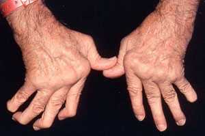
RA hands: symmetrical deforming arthropathy with synovial thickening.

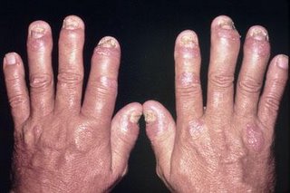
Psoriatic arthropathy: asymmetrical arthropathy involving terminal IPJ with nail changes.


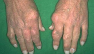
Chronic tophaceous gout: asymmetrical deforming arthropathy with tophi formation.

marfan syndrome

Friday, July 18, 2008

thumb sign

thumb sign ( ask patient to clench her thumb in her fist, the thumb should not exceed the ulnar side of the hand in normal subjects)

acromegaly hand


PACES exam, i got an acromegaly case. The stem being " please examine the patient's hands".

By one look at the patient, we are happy to conclude that the patient has acromegaly, the next thing in my mind is what the examiner want me to pick up from the patient's hands with acromegaly.

Possible things to pick up:
1) Carpal Tunnel syndrome
2) Osteoarthritis
3) Pure hand feature of acromegaly

We may easily get a 4 mark if we follow the stem, go accordingly to point out the features that support the diagnosis.
Steps to follow in order for acromegaly patient in the very moment of 2 min before we use up 3 min for presentation.
1) Hand : large spade like hand, doughy hand, sweaty palm, tinel's sign

2) armpit: especially to look for skin tag (most candidate miss it, and we may only get 3 if we miss it)

3) Face: Prominent supraorbital ridge, prognatism, widely-space teeth, macroglossia, increase in skin creases/ wrinkles

4) Visual field assessment -bitemporal hemianopia

5) Lower limb: large foot, osteoarthritis of knee, thicken heel pad

6) complete by mentioning: checking BP, urine for glycosuria, examine for galactoria

Present as such order to make it systematic but the opening sentence must be base on the stem, in this case, "examine the hand"
Eg:
This patient has features of acromegaly. I say so because she has a large doughy, spade like hand. Her palm appeared sweaty. There is evidence of carpal tunnel syndrome as the tinel sign is positive. I found a skin tag over her right axillary area. Her voice appears to be coarse, she has prominent supraorbital ridge, and prognatism. She has macroglossia with widely-space teeth. Her foot appears to be large. Her heel pad is thickened. There is sign of osteoarthritis over her knee joint. There is no evidence of bitemporal hemianopia.

In conclusion, Mdm X has acromegaly. The most likely cause is a macroadenoma. There is sign of active disease in view of presence of sweaty palm, and skin tag. I would like to further assess her cardiovascular system.

These are the questions the examiner asked from me: ( not much time left after my presentation)
1) How do you confirm the diagnosis
Answer: Failure of suppression of GH level to less than 2ng/ml after oral glucose confirm the diagnosis
2) What further test you want to do?
Answer: Localization of pituitary tumour by requesting a MRI of brain
3) What are the treatment?
optimization of cardiovascular risk by strict BP and sugar control, refer eye for Visual field perimetry, definitive treatment: transphenoidal hypophysectomy, medical: somatostatin analogue: Octreotide, GH receptor antagonist: Pegvisomant

That end the session with clear pass from both examiner.
Further possible question:
1) What other source of GH if MRI shows no macroadenoma?
Answer: ectopic growth hormone production, small cell CA lung
2) How do you monitor treatment response?
Answer: insulin like growth factor
3) Do you know of any endocrine adenoma association with acomegaly?
Answer: MEN type 1 -parathyroid hyperplasia, pituitary adenoma, pancreatic tumor
Need to examine calcium level

acromegaly disease is active or not, what is the full answer?

acromegaly disease is active or not, what is the full answer?


sweating, hypertension, glycosuria and headaches will indicate disease activity

Don't miss a scar in hypothyroid patient!

General inspection
** Hoarse voice, response and movements are slow
** Overweight
** Myxoedematous facies as shown in the picture

Check eye for jaundice(?chronic active hepatitis)and pallor (?hemolytic anemia). Don't forget to look for Grave's disease eye signs.

Rub the hair with your fingers to feel for coarse hair.

Check the mouth for macroglossia.

Check the neck for goitre and scar
Don't miss a scar in this case! In PACES, 99.9% of hypothroidism is due to previous total thyroidectomy.

Thyroidectomy scar!

Then, proceed to the hand to count the pulse rate i.e. bradycardia and look for acropachy in Grave's ds.

Test for proximal myopathy over shoulder.

Check for pretibial myxoedema and pedal edema in lower limbs.

Lastly, position the patient in a chair to test the ankle jerk for slow relaxation phase. Remember, this is the most important sign that indicate the patient is clinically hypothyroid!

End your examination by saying that you would complete it by checking CVS, Lung and cerebellar signs.

Learning points:
** Don't miss a scar in hypothyroid patient!
** Always look for Grave's ds signs in hypothroid patient. Remember, the treatment may cause hypothroidism!


Cushing's syndrome

Cushing's syndrome is another popular spot diagnoses in PACES. The candidate is usually asked to "Look at the patient's face".

There are 3 main steps in dealing with this case:

First, be able to recognise all the striking features of Cushing's syndrome.
** Typical moon-face with plethora, hirsutism and acne.
** Truncal obesity with interscapular and supraclavicular fad pads.
** Purple striae over the abdomen, around the shoulders and breasts and thighs.
** Thin skin and easy brusing commonly found over limbs
** Proximal myopathy (shoulders & hips) and spinal tenderness (osteoporosis)

Then, you must always look out for the possible underlying diagnosis as it is not enough for a PACES candidate to get the diagnosis of Cushing’s syndrome only in MRCP.

Lastly, please complete your examination by mentioning that you would like to measure the BP and test the urine for sugar.

Points to remember: The commonest cause is still iatrogenic i.e. secondary to steroid. So, please look for RA hands, gouty arthritis, nephrotic syndrome and etc.

cushing

turners

lobectomy

In upper lobectomy , the lower lobe fills up the space left in the upper lobe thereby pulling up the diaphragm up , hence a raised diaphragm on side of lobectomy and clinical signs ( dullness, reduced vocal resonance) are confined to lower chest zones. The findings are same in lower lobectomy

a scar is a gift!"

a scar is a gift!"
As in this case with a big thoracotomy scar, one could localize the lesion side by the presence of the scar. Then, you should pay attention to the air entry during auscultation.
a. If air entry is reduced, then think of lobectomy and pneumonectomy.
b. If air entry is equal, then think of decortication and bullectomy.

"Which side first?

"Which side first?"

One of the favourite question my friends ask me for respiratoryry station. It's really depend on what you gather from the peripheral examination namely the tracheal position. If the tracheal is deviated, then I would advise you to concentrate on anterior chest first especially the apical region looking for apical fibrosis or collapse.
If the tracheal is central, then ask the permission to proceed with the back. The differentials would include fibrosing alveolitis, bronchiectasis, pleural effusion or consolidation.

lobectomy vspneumonectomy.

The differences:
1. The signs of lobectomy are confined to lobe which is removed. The signs are similar to pleural effusion except it is not stony dull.

2. The signs of pneumonectomy are extensive i.e. involve the whole lung. The side involved would be flatten. It is similar to whole lung collapse.

3. Normally, the tracheal is central in lobectomy except for upper lobe. The tracheal is almost always shifted in pneumonectomy.


dDX of creps and clubbing -
ca. of lung, fibrosing alveolitis, lung abscess

cause for fibroising alveolitis

The respiratory case that I had in UK was rheumatoid lung disease. I got no problem in detecting the clubbing and fine inspiratory crepitations bibasally. But, the outcome was 2/2!

Pitfall: I failed to identify the underlying cause for fibrosing alveolitis. I missed out the obvious sign i.e. Rheumatoid hand changes!

The moral of the story to learn is ALWAYS look for the underlying cause for fibroising alveolitis as cryptogenic fibrosing alveolitis is RARE for PACES!

Face: malar rash (SLE)
telangiectasia, beak nose, microstomia (scleroderma)
facial pigmentation (amiodarone)
heliotrope rash (dermatomyositis)
Hand: RA, sclerodactyly

causes of gynaecomastia

Station 1 : Abdomen - Cirrhosis of liver

Station 1 - Resiratory - Bronchogenic carcinoma

Station 3 : Cardiovascular - Drugs (spironolactone/digoxin)

Station 3 : Neurology - Myotonic dystrophy

Station 5 : Endocrine - Klinefelter's syndrome

I know there are other causes of gynaecomastia

HELICON KIT:

HELICON KIT:
H.Pylori eradicating kit
Each Helicon Kit contains: One Lansoprazole INN 30mg Capsule. Two Amoxicillin BP 500 mg Capsules UPS 500 mg tablet.
Indications: Helicon Kit is indicated in the eradication of H. pylori in active chronic gastritis, duodenal and gastric ulcers.

Mannitol IV

Mannitol IV

Indications & Dosage

INDICATIONS

Mannitol I.V. (Mannitol Injection, USP) is indicated for the following purposes in adults and pediatric patients.

Therapeutic Use

1. Promotion of diuresis in the prevention or treatment of the oliguric phase of acute renal failure before irreversible renal failure becomes established.

2. Reduction of intracranial pressure and brain mass.

3. Reduction of high intraocular pressure when the pressure cannot be lowered by other means.

4. Promotion of urinary excretion of toxic materials.

Diagnostic Use

Measurement of glomerular filtration rate

Reduction of Intracranial Pressure and Brain Mass

Reduction of Intracranial Pressure and Brain Mass: In adults a dose of 0.25 to 2 g/kg body weight as a 15% to 25% solution administered over a period of 30 to 60 minutes; pediatric patients 1 to 2 g/kg body weight or 30 to 60 g/m2 body surface area over a period of 30 to 60 minutes. In small or debilitated patients, a dose of 500 mg/kg may be sufficient. Careful evaluation must be made of the circulatory and renal reserve prior to and during administration of mannitol at the higher doses and rapid infusion rates. Careful attention must be paid to fluid and electrolyte balance, body weight, and total input and output before and after infusion of mannitol. Evidence of reduced cerebral spinal fluid pressure must be observed within 15 minutes after starting infusion

Thursday, July 17, 2008

efficacy and safety of albendazole

This comprehensive review briefly describes the history and pharmacology of albendazole as an anthelminthic drug and presents detailed summaries of the efficacy and safety of albendazole's use as an anthelminthic in humans. Cure rates and % egg reduction rates are presented from studies published through March 1998 both for the recommended single dose of 400mg for hookworm (separately for Necator americanus and Ancylostoma duodenale when possible), Ascaris lumbricoides, Trichuris trichiura, and Enterobius vermicularis and, in separate tables, for doses other than a single dose of 400mg. Overall cure rates are also presented separately for studies involving only children 2–15 years. Similar tables are also provided for the recommended dose of 400mg per day for 3 days in Strongyloides stercoralis, Taenia spp. and Hymenolepis nana infections and separately for other dose regimens. The remarkable safety record involving more than several hundred million patient exposures over a 20 year period is also documented, both with data on adverse experiences occurring in clinical trials and with those in the published literature and/or spontaneously reported to the company. The incidence of side effects reported in the published literature is very low, with only gastrointestinal side effects occurring with an overall frequency of just >1%. Albendazole's unique broad-spectrum activity is exemplified in the overall cure rates calculated from studies employing the recommended doses for hookworm (78% in 68 studies: 92% for A. duodenale in 23 studies and 75% for N. americanus in 30 studies), A. lumbricoides (95% in 64 studies), T. trichiura (48% in 57 studies), E. vermicularis (98% in 27 studies), S. stercoralis (62% in 19 studies), H. nana (68% in 11 studies), and Taenia spp. (85% in 7 studies). The facts that albendazole is safe and easy to administer, both in treatment of individuals and in treatment of whole communities where it has been given by paramedical and nonmedical personnel, have enabled its use to improve general community health, including the improved nutrition and development of children.

svc obstruction

There is prominent veins over this patient's anterior chest wall. They are tortuous and dilated. The neck veins are engorged with loss of pulsation. The face looks plethoric and suffused.

This is superior vena cava obstruction.

This patient has undergone recent radiotherapy for this medical emergency condition as evidenced by fresh radiation ink marks and skin erythema over the same area.

"pink puffers"

Emphysema patients are sometimes referred to as "pink puffers". This is because emphysema sufferers may hyperventilate to maintain adequate blood oxygen levels. Hyperventilation explains why emphysema patients do not appear cyanotic as chronic bronchitis (another COPD disorder) sufferers often do; hence they are "pink" puffers (adequate oxygen levels in the blood) and not "blue" bloaters (cyanosis; inadequate oxygen in the blood).

Blue Bloater

stion: What is a Blue Bloater? What is Type B COPD?

Chronic Obstructive Pulomonary Disease (COPD) patients have both chronic bronchitis and emphysema. However, a patient will typically be classified as either suffering primarily from one or the other. Patients suffering primarily from chronic bronchitis are referred to as "blue bloaters", while patients suffering primarily from emphysema are referred to as "pink puffers".

Answer: The term "blue bloater" is derived from the bluish color of the lips and skin commonly seen in patients suffering from Type B Chronic Obstructive Pulmonary Disease (COPD).

Signs and Symptoms of Blue Bloaters

A blue bloater has a history of cough with sputum for 3 months to one year or more. A blue bloater experiences cyanosis due to a decrease in sufficient amounts of oxygen reaching the blood. Ankles and legs may be swollen and distention in the neck veins may be apparent. A blue bloater primarily suffers from chronic bronchitis. Blue bloaters develop signs of right-sided heart failure.

Pink Puffer

Question: What is a Pink Puffer? What is Type A COPD?

Chronic Obstructive Pulomonary Disease (COPD) patients have both chronic bronchitis and emphysema. However, a patient will typically be classified as either suffering primarily from one or the other. Patients suffering primarily from emphysema are referred to as "pink puffers", while patients suffering primarily from chronic bronchitis are referred to as "blue bloaters".

Answer: The term "pink puffer" is derived from the reddish complexion and the "puffing" (hyperventilation) seen in patients suffering from Type A Chronic Obstructive Pulmonary Disease (COPD).

causes of copd

Smoking

The primary risk factor for COPD is chronic tobacco smoking. In the United States, 80 to 90% of cases of COPD are due to smoking.[9] Exposure to cigarette smoke is measured in pack-years, the average number of packages of cigarettes smoked daily multiplied by the number of years of smoking. Not all smokers will develop COPD, but continuous smokers have at least a 25% risk after 25 years.[10] The likelihood of developing COPD increases with increasing age as the cumulative smoke exposure increases. Inhaling the smoke from other peoples' cigarettes (passive smoking) can lead to impaired lung growth and could be a cause of COPD. It should be noted that, although tobacco smoking is the most common cause of COPD, the inhalation or smoking of other substances or drugs, for example cannabis, are also linked to the development of COPD.

Intense and prolonged exposure to workplace dusts found in coal mining, gold mining, and the cotton textile industry and chemicals such as cadmium, isocyanates, and fumes from welding have been implicated in the development of airflow obstruction, even in nonsmokers[11]. Workers who smoke and are exposed to these particles and gases are even more likely to develop COPD. Intense Silica dust exposure causes silicosis, a restrictive lung disease distinct from COPD however less intense silica dust exposures have been linked to a COPD-like condition[12]. The effect of occupational pollutants on the lungs appears to be substantially less important than the effect of cigarette smoking[13].

How can we show something in php?

Hi,
I am sohel once again. Today I will describe to u how can we show a value with php.
There are three way to write something with php.

1. echo
2. printf();
3

Describe:
1. we can write the any value of variable & even string through "echo" in php.
e.g.,
Output: It is working.
// suppose $v=20
Output: Your amount is: 20

However , If we use single quote ' ' then output will be such:
// suppose $v=20
Output: Your amount is: $v

2. print(), printf() are generally used for formatted output.


Output: Your amount is: 20
Your amount is: 20.00

3. This method is

Such as,
output: You are a great painter


So these are the processes for showing values through php.

OK. See u later on another topic.

diabetes insipidus

Central diabetes insipidus is caused by damage to the hypothalamus or pituitary gland as a result of:

  • Head injury
  • Infection
  • Surgery
  • Tumor

Nephrogenic DI involves a defect in the parts of the kidneys that reabsorb water back into the bloodstream. It occurs less often than central DI. Nephrogenic DI may occur as an inherited disorder in which male children receive the abnormal gene that causes the disease from their mothers.

Nephrogenic DI may also be caused by:

comment on hand

diagnose hand

Wednesday, July 16, 2008

Common deformities in rheumatoid arthritis

Common deformities in rheumatoid arthritis are the Boutonniere deformity (Hyperflexion at the proximal interphalangeal joint with hyperextension at the distal interphalangeal joint), swan neck deformity (Hyperextension at the proximal interphalangeal joint, hyperflexion at the distal interphalangeal joint). The thumb may develop a "Z-thumb" deformity with fixed flexion and subluxation at the metacarpophalangeal joint, and hyperextension at the IP joint.

conus medullaris

The conus medullaris is the terminal end of the spinal cord. It occurs near lumbar nerves 1 (L1) and 2 (L2). After the spinal cord terminates, the spinal nerves continue as dangling nerves called the cauda equina. The upper end of the conus medullaris is usually not well defined.

Stages of NHL

Stages of NHL

The various stages of NHL (the Ann Arbor staging classification, developed for Hodgkin's lymphomas) are based on how far the cancer has spread throughout and beyond the lymphatic system, and whether constitutional symptoms (fever, night sweats, or weight loss) are present.

Stage I
"Stage I" indicates that the cancer is located in a single region, usually one lymph node and the surrounding area. Stage I often will not have outward symptoms.
Stage II
"Stage II" indicates that the cancer is located in two separate regions, an affected lymph node or organ within the lymphatic system and a second affected area, and that both affected areas are confined to one side of the diaphragm - that is, both are above the diaphragm, or both are below the diaphragm.
Stage III
"Stage III" indicates that the cancer has spread to both sides of the diaphragm, including one organ or area near the lymph nodes or the spleen.
Stage IV
"Stage IV" indicates that the cancer has spread beyond the lymphatic system and involves one or more major organs, possibly including the bone marrow or skin.

The absence of constitutional symptoms is denoted by adding an "A" to the stage; the presence is denoted by adding a "B" to the stage (hence the name B symptoms).

Staging in non-Hodgkin's lymphomas is far less significant in determining therapy than it is in Hodgkin's disease.


management of steven johnson syndrome

This are outlines of management of steven johnson syndrome given by a dermatologist from Ipoh GH:

1) Vital Sign monitoring
2) Withhold Offending drug
3) Adequate hydration
4) Strict I/O chart
5) Assess disease severity daily
6) Oral lesion: NaHCO3 gurgle 4hrly
Syrup nystatin 6hrly
7) Crusted lip lesion: use liquid parafin soak with gauze apply to lips for 10min 4hrly to soften the crust, then slowly remove it
8) Body and lim lesions: KMno4 wash to raw area 2-3x per day, normal saline bath bd then apply gerila cream to raw area after KMno4, aqueous cream 2hrly

facial rash

should think of 3 DD when you encounter facial rash possibly dt connective tissue ds

1. SLE
2. Dermatomyositis
3. Mixed CT disease

diagnose the rash

drusen

asteroid hyalosis

crao

Diabetic retinopathy

Diabetic retinopathy is a condition that affects the retinal blood vessels in patients with diabetes mellitus. It can be classified into:

  • Background diabetic retinopathy
  • Preproliferative diabetic retinopathy
  • Proliferative diabetic retinopathy
  • Diabetic macular edema
Background and preproliferative diabetic retinopathy are also referred to as nonproliferative diabetic retinopathy.

Tuesday, July 15, 2008

leopard/lamb

antibiotic treatment of anaerobic infections

Serious infections may require hospitalization for treatment. Immediate antibiotic treatment of anaerobic infections is necessary. Laboratory testing may identify the bacteria causing the infection and also which antibiotic will work best. Every antibiotic does not work against all anaerobic bacteria but nearly all anaerobes are killed by chloramphenicol (Chloromycetin), metronidazole (Flagyl or Protostat), and imipenem (Primaxin). Other antibiotics which may be used are clindamycin (Cleocin) or cefoxitin (Mefoxin).

Aftermath of the bombings

At least 35 people have been killed and more than 50 injured in a double suicide bombing north of the Iraqi capital, Baghdad, army sources say.

Are there approved hospitals / centres at which I can obtain work experience / training for PACES?

The Colleges' Examinations Offices do not have a list of accredited or recognised centres for training and work experience. There is therefore no requirement that candidates train or work at approved hospitals for the MRCP(UK) examination. Candidates may wish to check with the General Professional Training Department that their experience meets the requirements for progression in their career after having completed the MRCP(UK) examinations.

What are the eligibility requirements for the PACES examination?

Candidates are required to have passed or gained exemption from the Part 1 and Part 2 Written examinations. In addition, candidates need two and a half years' work experience / training since graduation. Twelve months of this two and a half years must have been spent in a Senior House Officer, equivalent or higher grade in acute medicine posts in which there is a strong emphasis on responsibility for unselected emergency admissions and the continuing care of acute emergency medical patients. Candidates also need to have gained their twelve months of Senior House Officer acute medicine experience within five years of the PACES examination they are taking.

It is also presumed (as in the UK) that all doctors will have undertaken a pre-registration year / year at a house officer level or equivalent in which they would have gained 6 months experience in acute medicine. If this has not been the case then you would also need to obtain a further 6 months of acute experience.

Iran: Talks with US possible soon

Iran: Talks with US possible soon

Iranian President Mahmoud Ahmadinejad
Ahmadinejad's comments come at a time of growing tension

Iranian leader Mahmoud Ahmadinejad has said talks with the United States are possible in the near future, according the country's official news agency.

But he said that such talks would not happen at governmental level

An undercover BBC investigation has exposed how young African footballers are being conned out of thousands of dollars by Nigerian fraudsters.

The Grand Canyon at Band-e Amir
It is an area of outstanding natural beauty

It takes eight bone-shaking hours on a dirt track road to reach Afghanistan's first national park from the capital, but the beauty and serenity is worth crossing the world for.

Giant oilfield to raise Saudi output

By Andrew Walker
Business correspondent, BBC World Service, Khurais, Saudi Arabia

Workers at the Khurais oil field
Workers brave the heat to get the project finished in time

The desert sun is beating down. The temperature is 44 degrees Celsius. And yet there are men working in the baking heat.

Not all that energetically, perhaps, but they are working nonetheless. For them, there is no escaping on board the visiting air-conditioned bus. Instead, simple shelters provide scant, yet welcome, relief.

Welcome to Khurais in the Saudi Arabian desert, 160 kilometres east of the capital Riyadh.

It is described by the national oil company, Saudi Aramco, as the largest oilfield development in the history of the industry, containing 27 billion barrels of oil.

Breeding rate fall for rare bird

Slavonian grebe (Pic: RSPB Images)
Slavonian grebe are one of UK's most colourful birds

One of the UK's rarest birds is facing its worst breeding season on record, according to RSPB Scotland.

Slavonian grebe first nested in Scotland near Loch Ness, Inverness-shire, in 1909.

 
Loading...