The most important reason for performing joint fluid analysis is to rule out septic arthritis. Synovial fluid analysis can demonstrate local inflammatory response, infection and the presence of crystals. Synovial fluid effusions are classified into five general etiologic categories: noninflammatory responses, inflammation, sepsis, crystal induced and hemorrhage. The most common diseases associated with each category are summarized in the following table.
The following tests can be performed on synovial fluid.
Synovial fluid protein concentration is usually 25% of serum (1-3 g/dL). Synovial fluid glucose is normally within 10 mg/dL of plasma glucose. Neither synovial fluid glucose nor protein provides much useful diagnostic information. White cell counts (WBC) are normally less than 200/uL with fewer than 25% neutrophils. A WBC count of 2,000/uL and a neutrophil count of 75% serve as useful cutoff points to distinguish inflammatory from noninflammatory disease. However, there is much overlap in synovial fluid WBCs between the inflammatory, crystal-induced, and sepsis categories. WBC is greater than 50,000/uL in 70% of patients with septic arthritis, 15% with gout, 10% with pseudogout, and 4% with rheumatoid arthritis. Acute septic arthritis is usually caused by bacterial infection of the joint. Tuberculosis and fungi are less commonly involved. Infection can occur by hematogenous spread, direct innoculation during surgery or trauma or from osteomyelitis. Septic arthritis is usually characterized by an acute onset of monoarticular joint pain. Staph aureus is responsible for the majority of cases of bacterial infection. Neisseria gonorrhea is common in adolescents and young adults. E. coli is most commonly seen in neonates. Other gram positive rods are associated with immunocompromised patients. Specimen requirement for cell count and differential is 1 mL of synovial fluid transferred to a lavender top (EDTA) tube. Bacterial cultures require 2-3 mL of fluid in a yellow top tube. | |||||||||||||||||||||||||||||||||||||||||||||||||||||||||||||||||||||||||||||||||||||||||||
 | |||||||||||||||||||||||||||||||||||||||||||||||||||||||||||||||||||||||||||||||||||||||||||
Sunday, July 20, 2008
Synovial Fluid Analysis


mrcp,rheumatology
mrcp,rheumatology
On examination, his temperature is 38.5°C. His eyes are red. His right knee is hot, swollen and tender to palpate. No other joint appears to be affected.
Investigations:
Hb 12.9 g/dl
WBC 14.0 x 10⁹/l
Platelets 200 x 109/l
ESR 75 mm/h
Blood cultures
No growth after 48 hours
Urinalysis
No blood, glucose or protein detected
Knee x-ray
Soft tissue swelling around left knee
What is the most likely diagnosis?
1 )Gout
2 )Gonococcal arthritis
3 )Reiter's syndrome
4 )Rheumatoid arthritis
5 )Viral arthritis
erythema nodosum,mrcp


Very frequent MRCP exam question, especially Part 1 & 2a, don't be surprised if it comes out in Station 5 PACES.
The picture shows erythematous nodules over the legs involving the shin. They are tender and elevated.
My diagnosis is that this patient has erythema nodosum.
You need to know the causes !
May even appear in history taking if you read Mir &Ryder PACES
Idiopathic/ unknown
Sarcoidosis
Streptococcal infection
Tuberculosis
Infections other than TB or strep - HIV...
Pregnancy or oral contraceptives
Drugs other than OCAs
Inflammatory bowel disease
Behcet's disease
Other
massive splenomegaly.+mrcp
Box 1 Diseases that cause massive splenomegaly.
- Chronic myeloid leukemia
- Extrahepatic portal vein obstruction
- Gaucher's disease
- Hairy-cell leukemia
- Idiopathic portal hypertension
- Malaria
- Malignant lymphoma
- Myelofibrosis
- Thalassemia major
- Visceral leishmaniasis (i.e. kala azar)
sarcoidosis in mrcp
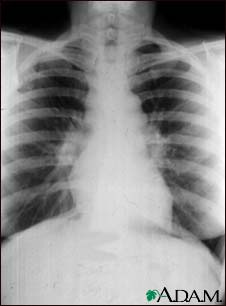
Remember that 90% of CXRs have bilateral hilar LN enlargement. More advanced case may have diffuse fibrosis!
Tips for MRCP
1) If you are asked about a patient with painful skin lesions over shin area with dry cough, always think of sarcoidosis.
2) A patient with high calcium level and dry cough , think of sarcoidosis although there is possibility of lung cancer with bone metastasis!
- Stage 0 is a normal chest x-ray.
- Stage 1 is a chest x-ray with enlarged lymph nodes but otherwise clear lungs.
- Stage 2 is a chest x-ray with enlarged lymph nodes plus
infiltrates (shadows) in the lungs. - Stage 3 is a chest x-ray that shows the infiltrates (shadows) are present but the lymph nodes are no longer seen.
- Stage 4 shows scar tissue in the lung tissue.
Saturday, July 19, 2008
tuberculous arthritis
Unlike tuberculous osteomyelitis, which typically involved the thoracic and lumbar spine (50 percent of cases), tuberculous arthritis primarily involves the large weight-bearing joints, in particular the hips, knees, and ankles, and only occasionally involves smaller non weight bearing joints.
Progressive monarticular swelling and pain and develop over months to years, and systemic symptoms are seen in only half of all cases. Coexistent active pulmonary tuberculosis is unusual.
Aspiration of involved joint yields fluid with an average cell count of 20,000/µL, with approximately 50 percent neutrophils. Acid fast staining of the fluid yields positive results in fewer than one-third of cases, and cultures are positive in 80 percent.
Mercedes Benz scar
Mercedes Benz scar

This is the coolest thing I learned about today. The Mercedes Benz scar indicates previous liver transplant. In the patient we saw the lower parts of the sacar did not have an angle so it looked more like two scars, a transverse with a midline ie: like the mathematical sign for parallel. I get excited by scars I don't know about as you might have already realised :)) I think it is because I like patterns... and also this one is particularly exciting because of the name- it hasn't been long since I used to read the car brand as the French for Wednesday (Mecredi), so the mention of Mercedes makes me laugh at my dyslexic tendencies :)

 Posts
Posts


